Educated Patient® Lung Cancer Summit Advancements in Diagnosis: April 29, 2023
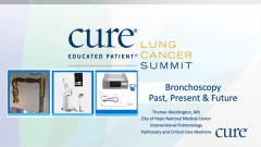
Watch Dr. Thomas Waddington, from City of Hope National Medical Center, discuss the past and future of bronchoscopy during the CURE® Educated Patient® Lung Cancer Summit.
Episodes in this series

Detection of lung cancer with bronchoscopies has advanced over the past few years, allowing providers to stage tumors and potentially determine treatment options, one expert said.
Dr. Thomas Waddington, director of interventional pulmonary medicine at City of Hope National Medical Center in Duarte, California, discussed the progression of advancements in this space during the CURE® Educated Patient® Lung Cancer Summit.
“I think what I was trying to get across as much as anything else is how much easier (bronchoscopy) is now as compared to where it was in the past, (and) how much safer it is now as compared to what it was in the past,” he said during an interview with CURE®. “And when you diagnose a nodule, you’re also supposed to stage a nodule and also figure out how to treat it. The best way to do that is all in one procedure, (and) we can do that now.”
According to the National Cancer Institute, a bronchoscopy is a procedure performed to look inside the trachea, air passages and lungs via the nose or mouth to detect cancer or perform some treatment procedures.
Waddington told CURE® that most patients with suspected lung cancer are physically able to undergo bronchoscopy.
“Right now, honestly, unless the patient is severely debilitated, this is still, by far, the safest and easiest procedure for them to go through,” he said. “The only patient I would actually say cannot (undergo bronchoscopy) is one where the anatomy is just too distorted from the tumor to make it possible and/or the patient decides they just don’t want to go through the procedure because they’re not looking into active treatment.
Where It Began
The first bronchoscopy was performed in 1876, and since then, it has advanced with tools making the procedure easier to perform and more accurate.
For example, a prototype of a flexible bronchoscope was developed in 1964, then another flexible bronchoscope was presented in 1966, leading to its commercial availability in 1968.
“The flexible bronchoscope was really where bronchoscopy and pulmonology became a much more integral field in cancer and diagnostics,” Waddington said. “That was a big change because we went from this therapeutic intervention, removing foreign bodies, to a much more diagnostic approach in the field.”
Ultrasound technology was introduced to bronchoscopy in the 1990s, which “completely changed my field,” Waddington said. This allowed health care providers to tell the difference between vessels, lung tissues and cancers. Ultrasound technology was added to the end of a bronchoscope itself in 2004.
“That completely changed what we did because now, bronchoscopists could look between lungs at lymph nodes, and this is often where cancers or other diseases will travel to,” Waddington said.
Electromagnetic
Improving Limited Accuracy
Despite all of these advances in bronchoscopy, Waddington said that the accuracy of these approaches was only around 70%. Interventional radiologists were able to access these nodules with 92% accuracy, even though that involved minimally invasive procedures. In addition, airways including the lung cover approximately 142 meters square of surface area, which Waddington compared to trying to find a dime in a three-bedroom house.
Approximately 70% of lung cancer nodules are located in the outer third of the lung, and this led to the
Future Advancements
Waddington said that he believes the future is going to entail a combination of endobronchial ultrasound, robotic technology and even local therapy like thermal ablation, chemotherapeutics and immunotherapies.
“We’re going to use these things to be able to cure people and create survivors,” he said.
One example of integrating bronchoscopy with therapies is photodynamic therapy. This involves taking a photosensitizing agent — which Waddington said was similar to sun exposure at the beach — and inject it into the patient, where it concentrates into tumors.
Pulsed electric field energy is another potential therapy on the horizon and involves non-thermal therapy that applies high-frequency biphasic electric pulses placed into a tumor, Waddington explained. This destabilizes the membrane of the tumor, allowing tumor fragments to be released into the bloodstream.
This new technology, which was released a few months ago, has demonstrated positive results in studies presented at recent medical conferences. One study’s findings demonstrated that patients treated with pulsed electric field energy had no heart-related side effects and they had minimal scar tissue after treatment.
“Over the last 15 to 20 years, what we've noticed is chemotherapy is a very toxic agent (but) incredibly useful drugs,” Waddington said. “But if we can activate your immune system to go recognize a tumor and destroy it, it's incredibly effective. It lasts a whole lot longer. And this pulse electric field (energy) is allowing the body to start to do that.”
For more news on cancer updates, research and education, don’t forget to


